Unidentified Arthropods in the Christopher B. Smith Preserve
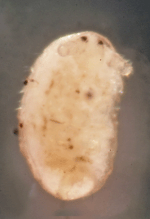
Arthropod Characteristics: All arthropods are invertebrate animals with exoskeletons (external skeletons), segmented bodies, and jointed, paired appendages. As an arthropod grows, it molts its exoskeleton.
Arthropods include members of Class Arachnida (spiders and mites), Class Branchiopods (water fleas), Class Malacostraca (shrimp and wood lice), Class Diplura (diplurans), Class Collembola (springtails), Class Insecta (insects), Class Chilopoda (centipedes), Class Diplopoda (millipedes), and Class Symphyla (dwarf millipedes and garden centipedes).
There are so many described species of arthropods in the world that they make up more than 80% of all described living animal species.
Interactions in the Smith Preserve: Depending on the species, they are pollinators, predators, and prey. Some help recycle nutrients in the soil, while others help transmit diseases to plants and animals.
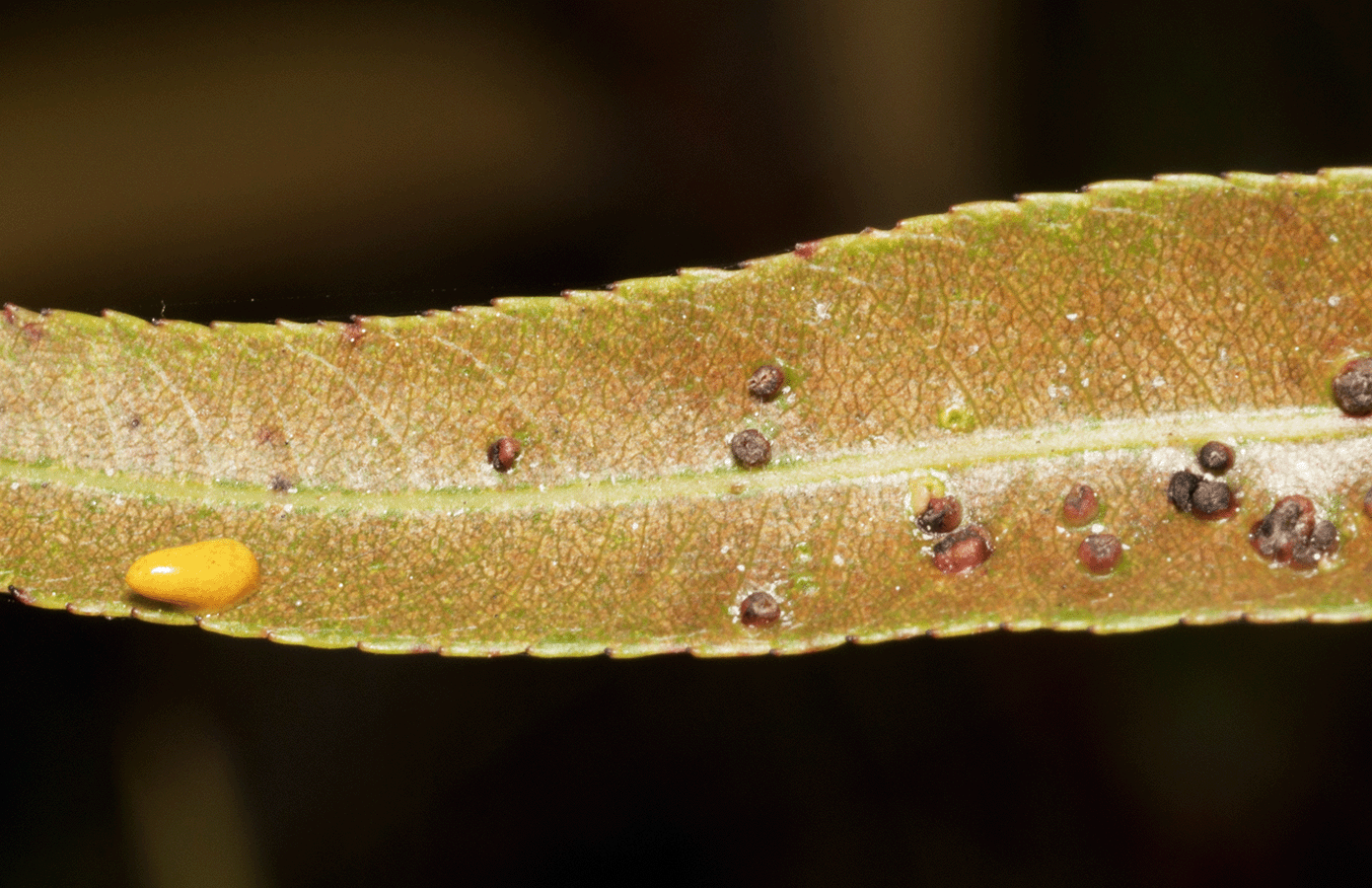
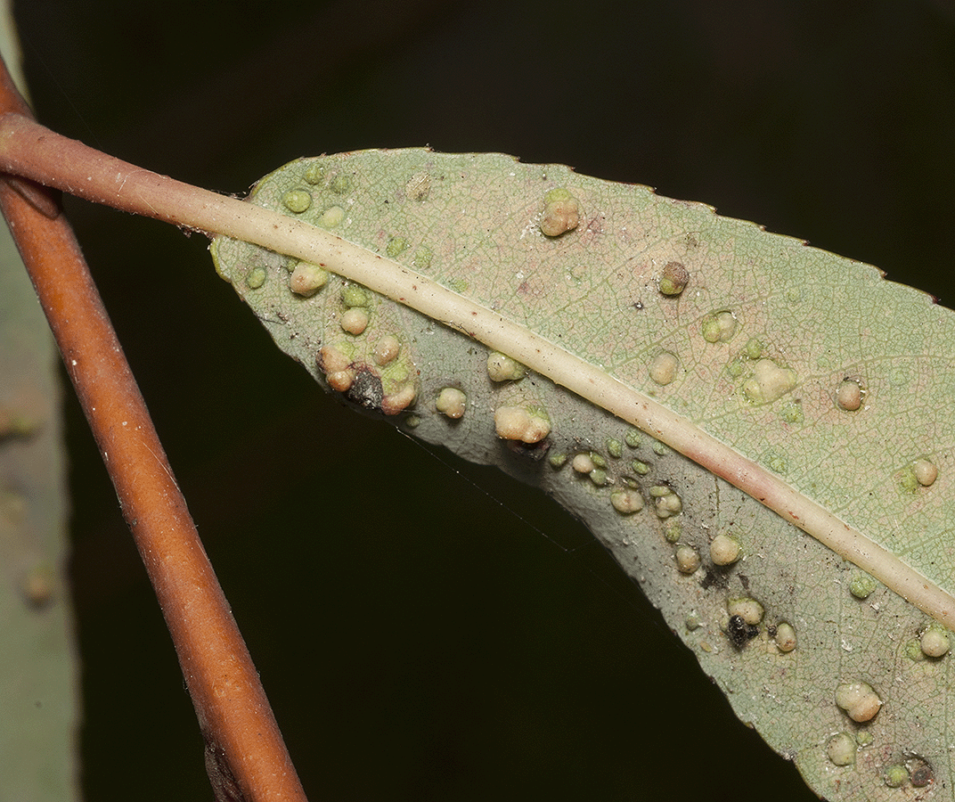
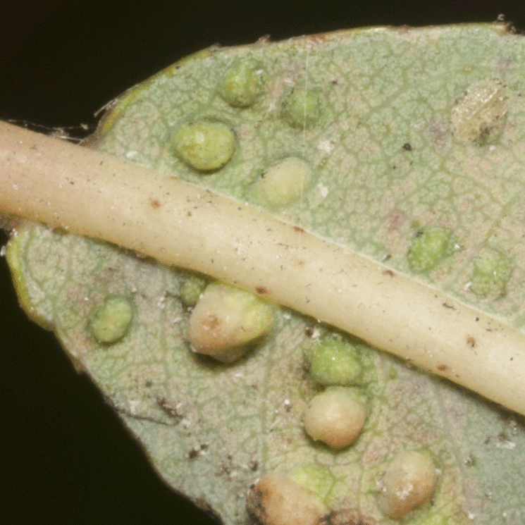
Special Note: The photographs and descriptions of the arthropods on this web page have been submitted for identification to the experts at <BugGuide.net>. To date, none have had a confirmed identification, even to class.
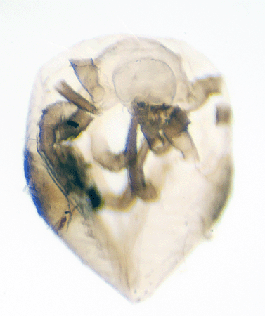
Unknown Arthropod #1
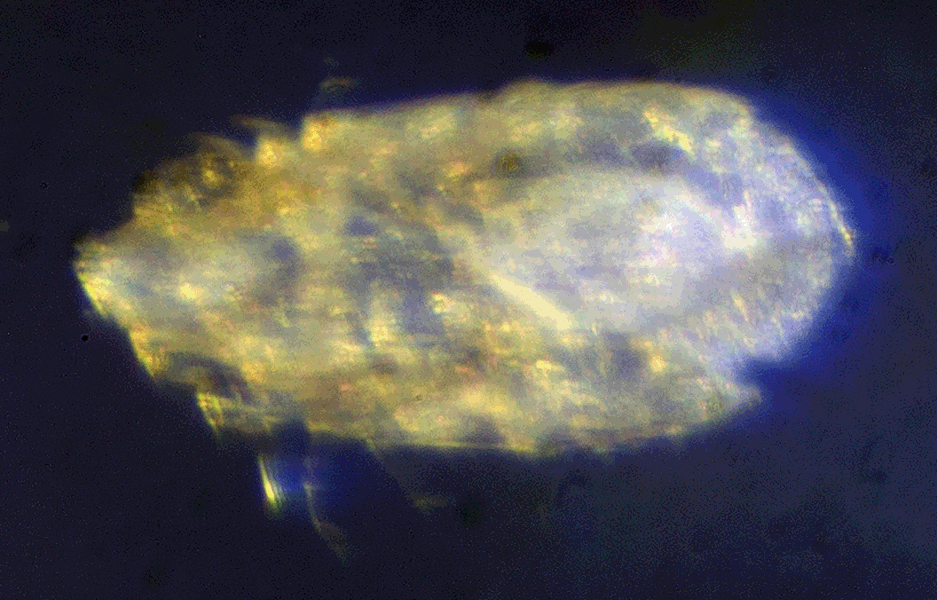
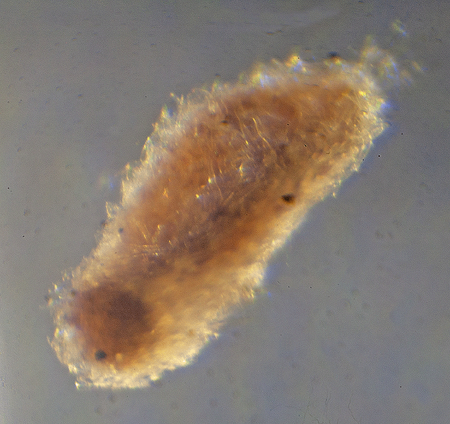
This 2.75 mm arthropod was living in leaf litter beneath an oak hammock north of the Smith Preserve Way bridge on February 10, 2016.
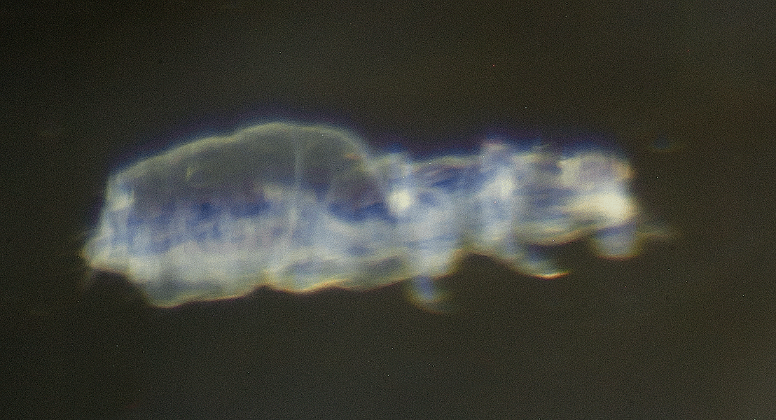
The individual was isolated from the litter by using a Berlese Funnel, and these photographs were created using photomicroscopy. The photographs were submitted for identification to <BugGuide.net> on 13 March 2016. To date, no one at <BugGuide.net> has identified this animal.
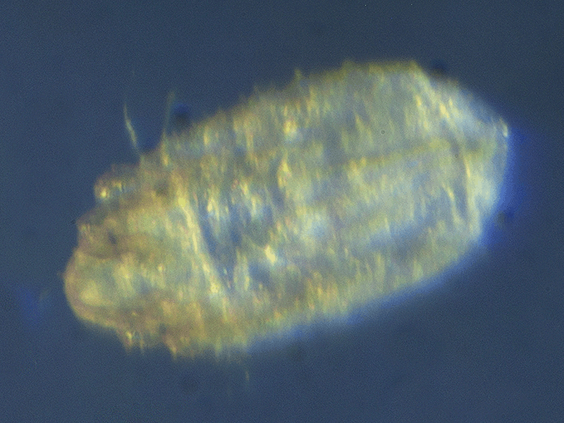
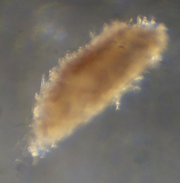
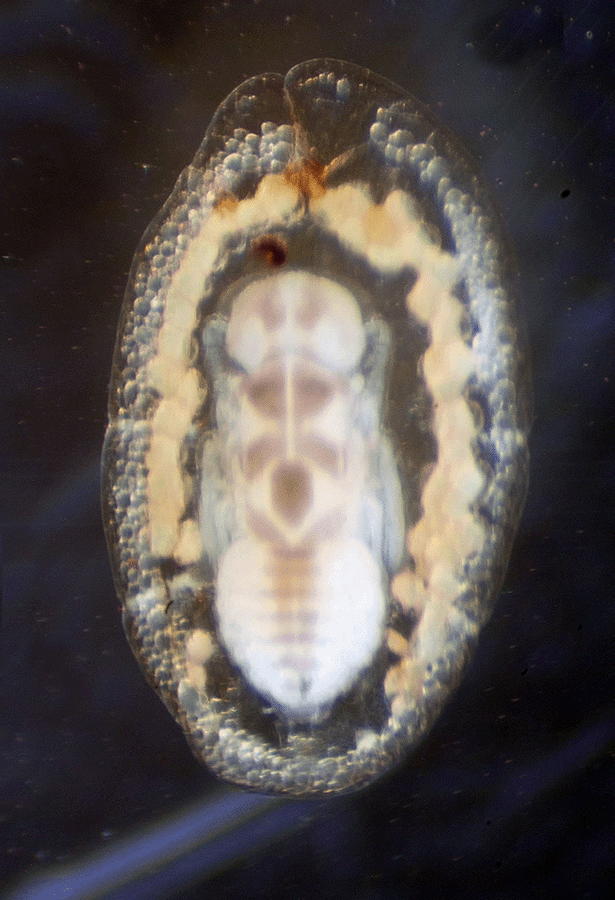
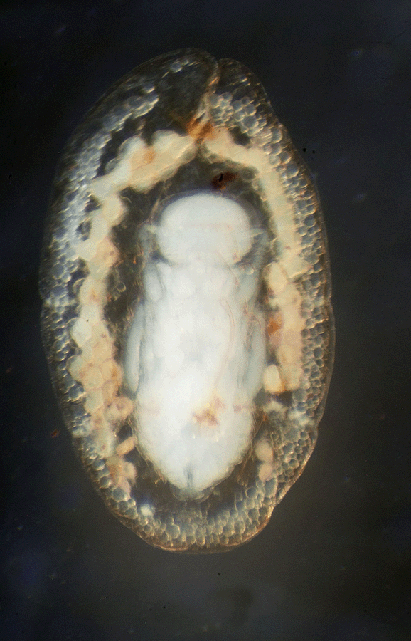
The 3.75 mm long arthropod below was found in the same leaf litter sample as the individual above. The process for removing this specimen and photographing these images is the same as above.
The first photograph below is a dorsal view and the second, a ventral view. As can be seen in the ventral view below, the dorsal "capsule" seemed to wrap around the legs. This characteristic was also true of the specimen above.
These two photographs were submitted to <BugGuide.net> for identification on 4 March 2016. This specimen, like the one above, has yet to be identified. It is likely the same species, but this one is 1 mm larger than the one above.
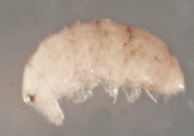
Unknown Arthropod #2
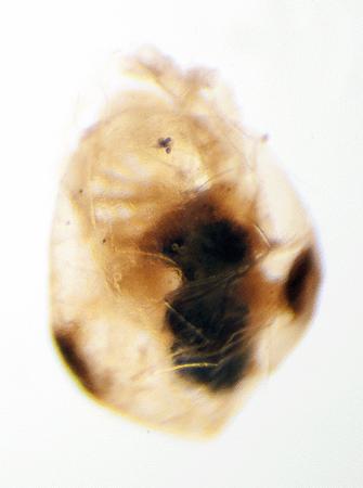
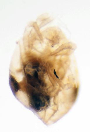
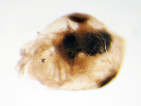
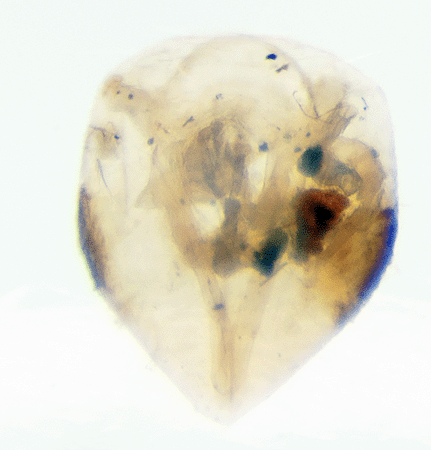
This specimen, .75 mm in length, was found in wet pine litter in the southeastern corner of the Smith Preserve on 9 December 2015. The litter sample was collected after it had rained for two consecutive days.
The specimen was extracted from the litter with a Berlese funnel and these photographs were created using photomicroscopy. The first image is a lateral view, the second is a dorsal view, and the third and fourth are ventral views.
The specimen appears to have distant segments along its dorsal surface. Its legs are bulbous and there do not appear to be segments. There are more than 3 pairs of legs. Some legs appear to have claws.
The dorsal surface is curled over and resembles a mm poly. There are what look like chopped off antennae as well as some kind of extended mouth structure. There are distinctive eyes/eyespots with hairs nearby that look almost like eyelashes.
Although these images were submitted to <BugGuide.net> on 22 December 2015, the scientific class of arthropod has yet to be identified.
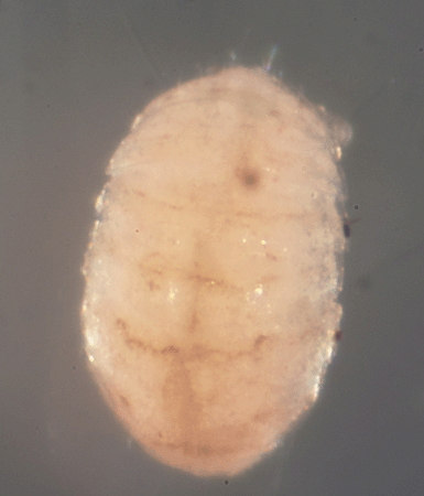
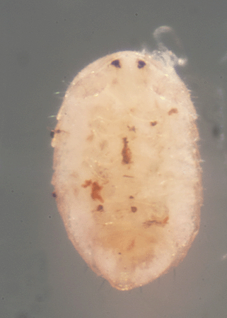
Unknown Arthropod #3
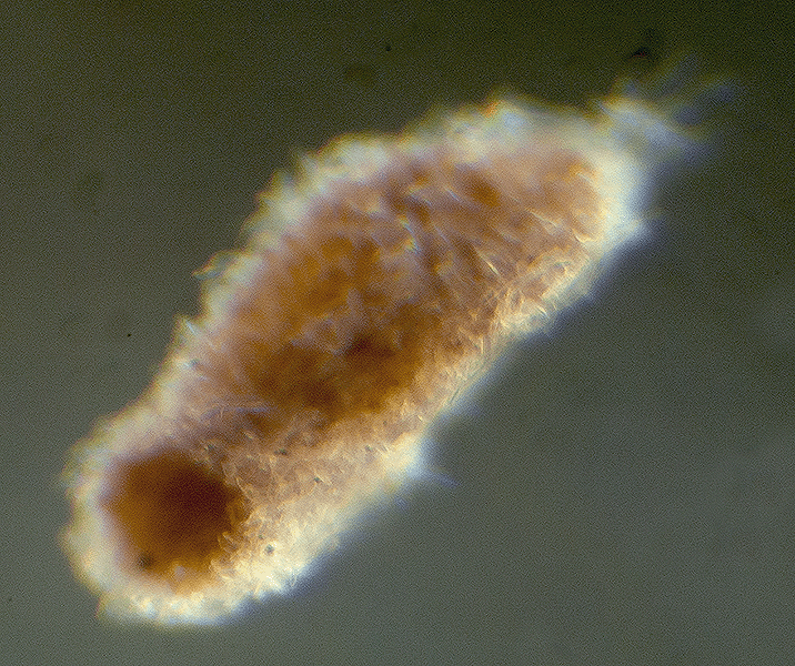
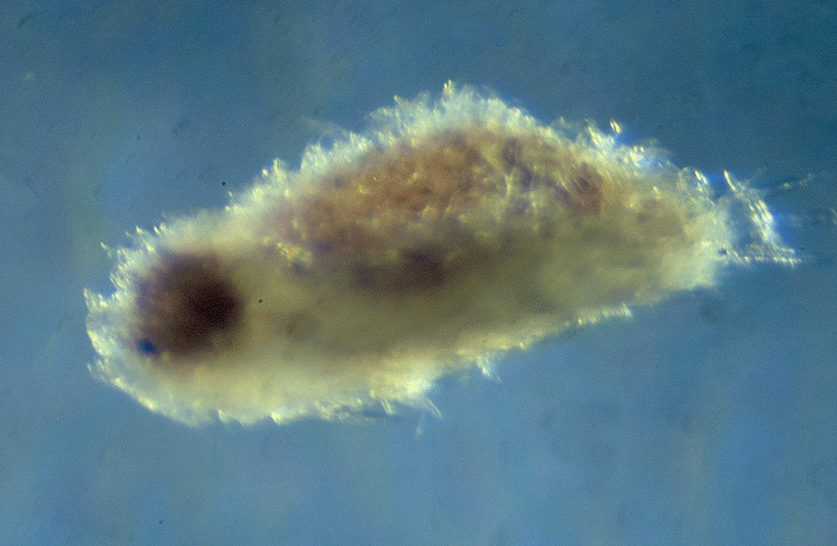
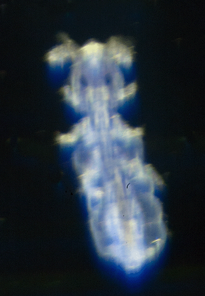
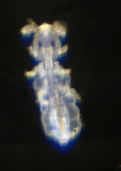
This .2 mm long arthropod was living in leaf litter collected beneath a Serenoa repens (Saw Palmetto) bush and a Quercus geminata (Sand Live Oak) tree on January 3, 2017, just north of Smith Preserve Way and to the right of the center of this part of the preserve.
The individual was isolated from the litter with a Berlese funnel and these images were created using photomicroscopy. The images were sent for identification to <Bugguide.net>. To date, no one has helped with the identification.
This individual appears to have eyes and is very flattened. The head appears to be on the left. The webmaster believes the first image is a dorsal image and the second is a ventral image. The second image has two appendages folded over the body from what is thought to be the anterior end. Are these appendages, mouth structures, antenna, legs, or something else?
Unknown Arthropod #4
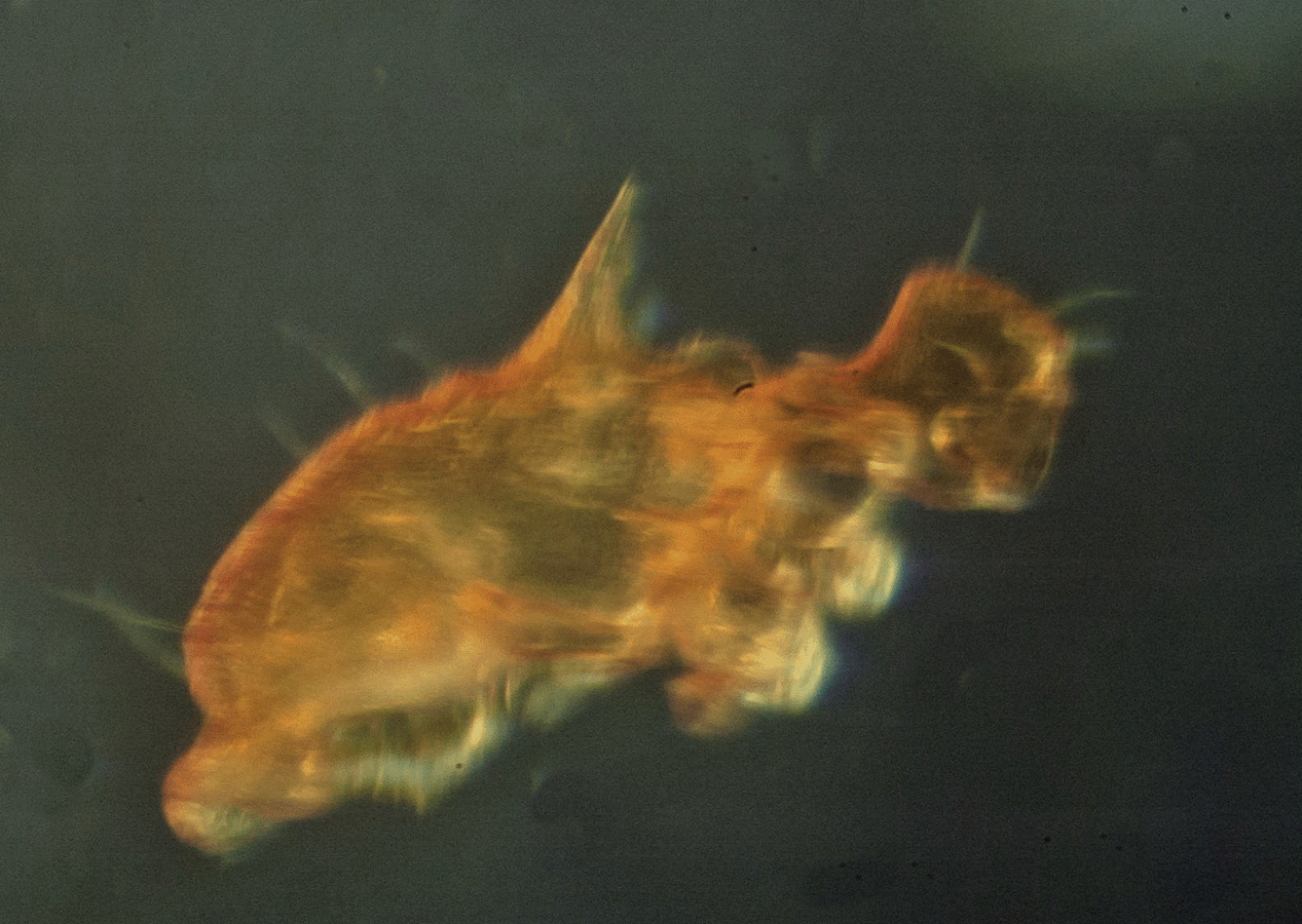
On November 16, 2022, this .8 mm spiny individual was captured in a pitfall trap that had been left overnight in a hammock in the northeast quadrant of Smith Preserve. These images were created by photomicroscopy and sent for identification to <Bugguide.net>, sponsored by Iowa State University's Department of Entomology. To date there has been no identification of this species.
It appears to have a dark head with two tiny eyes. The last image is a ventral shot.
Unknown Arthropod #5
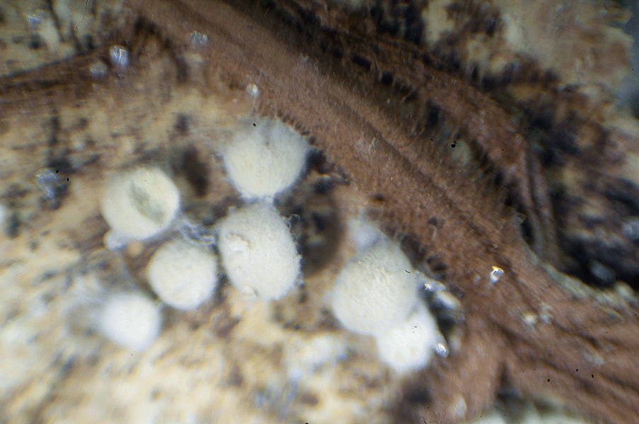
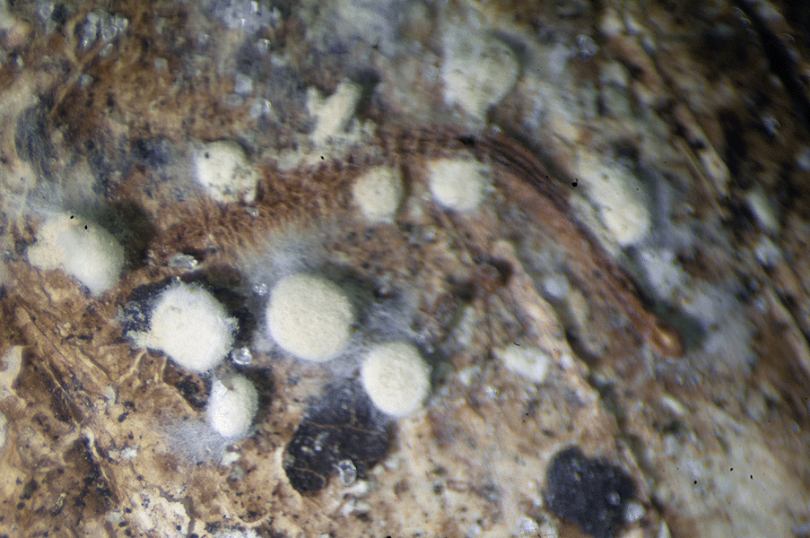
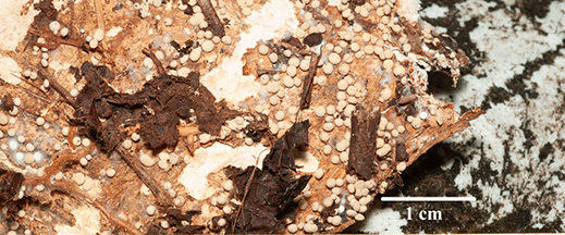
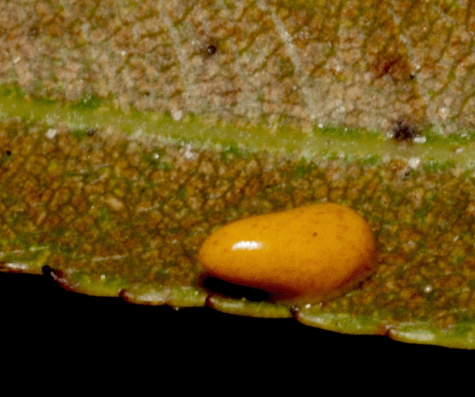
On January 31, 2023, scutes (hard scales making up the shell that look like separate plates) from a deceased gopher tortoise were examined in the Smith Preserve near 14th Avenue. On the bottom of several scutes were what appeared to be white eggs and/or globular-shaped fungus colonies.
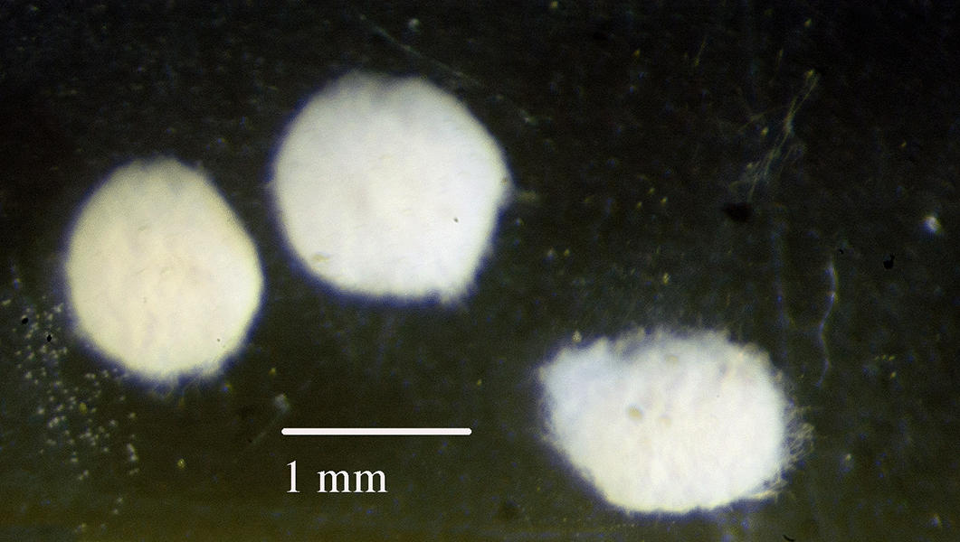
The first photograph below shows what looks one or more dented eggs that may have hatched. In closer photos, the "eggs" appear to have insect silk or fungal mycelial mats around them. (Mycelial mats are root-like structures of a fungus consisting of a mass of branching thread-like hyphae. Hyphae are the branching filaments that make up the mycelium of a fungus.)
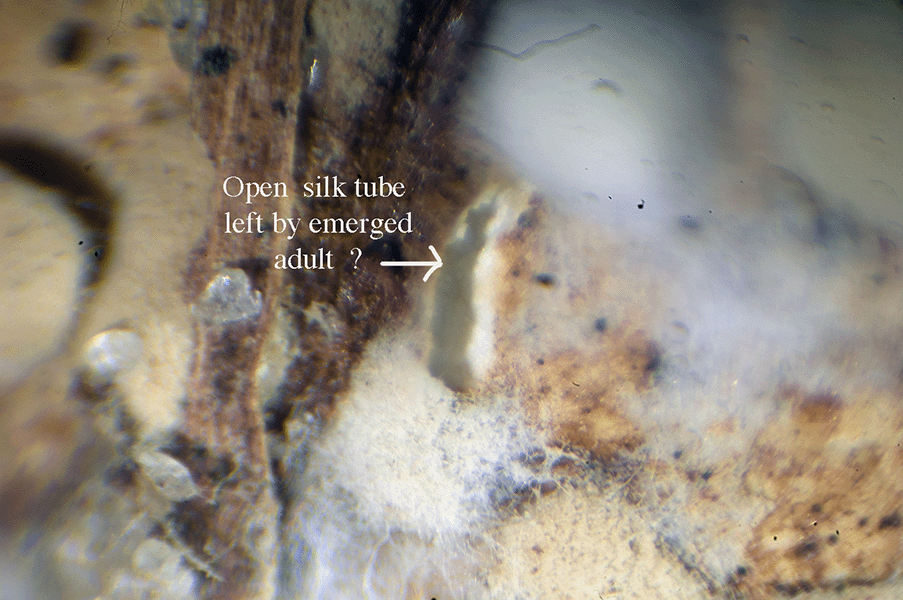
In the image below, it looks like a caterpillar may have emerged from a silk cocoon.
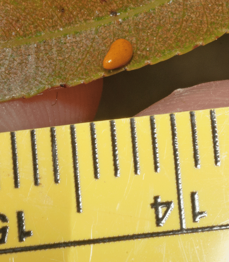
The two images below show more of these rounded objects. The last photo shows that these round objects are approximately 1 mm in diameter.
Images have been sent for identification to <Bugguide.net>, sponsored by Iowa State University's Department of Entomology; The Mushroom Observer, an online fungus identification group; and to Dr. Mark Deyrup, Emeritus Research Biologist at Archbold Biological Station, Venus, Fl. In 2005, Dr. Deyrup discovered a gopher tortoise tineid moth (Ceratophaga vicinella) that feeds on the keratin-rich shells of gopher tortoises. The webmaster is curious whether these round spheres could be eggs of the moth. To date, there has been no identification made by any expert.
Images were sent to iNaturalist and one contributer to iNaturalist suggested they were slime molds belonging to Phylum Mycetozoa.
Unknown Arthropod #6
On February 14, 2023, this 0.5 mm long arthropod was isolated from a dead Tillandsia fasciculata that had fallen from a tree, growing in the southeast quadrant of the Smith Preserve, not far from the east gopher tortoise fence.
These images (both ventral views above) were created using photomicroscopy and sent for identification to <Bugguide.net>, sponsored by Iowa State University's Department of Entomology.
The webmaster thought this might be a collembolan, but that identification was rejected by Frans Janssens on April 23, 2023. Janssens is a Contributer to <Bugguide.net> and is with the University Department of Entomology, Department of Biology, University of Antwerp, Antwerp, B-2020, Belgium. He is an authority on collembola: Bellinger, P.F., Christiansen, K.A. & Janssens, F. 1996-2014. Checklist of the Collembola of the World.<http://www.collembola.org>.
Below is a lateral view.
Unknown Arthropod #7
On February 17, 2024, the arthropod below, which appears to be inside an egg, was captured with a sweep net used in Ceratiola ericoides (Florida Rosemary) bushes. The total length of the object, with its surrounding capsule, was 1.25 mm. These images were created using photomicroscopy, and sent for identification to <Bugguide.net>, sponsored by Iowa State University's Department of Entomology.
When I submitted these images to <Bugguide.net>, I thought the first image was the ventral view with the hemipterian - like straw mouth part extending from the head. I thought the second image was the dorsal view. The antennae are at the top and appeared to be to be connected to the dorsal portion of the round head.
On January 6, 2025, the following response was received from John van der Linden, a Contributing Editor of <Bugguide.net>: "Hi Susan
I spent a while trying to track down what this might be, but couldn't find a clear answer. I do think it is probably something in Hemiptera or Psocodea.The outer, transparent, oval case looks most like the covering of a scale insect or a whitefly pupa. More like a scale insect since I believe whitefly pupae are usually a bit less elongate and more neatly symmetrical. The insect inside looks like an (unidentified) pupa to me. In contrast to your suggestion, I would posit that the current image [my first image above] shows the dorsal side of the pupa and the second image the ventral side; a close look at the second image reveals what appear to be the tips of the antennae and bases of legs, which you'd expect on the underside of a pupa; plus it seems more likely that the pigmented side of the animal (current image) would be the upperside.
If we assume the outer covering is the work of a whitefly or scale insect, there are a few possibilities. The insect inside could be the successfully developing pupa of the whitefly or scale insect itself, but I think this is unlikely -- I don't think adult scales or whiteflies usually have this kind of dorsal pigmentation, and also, the fact that the pupa doesn't fill the casing and is tilted at an angle suggests it's not the creator of the outer covering.
The remaining possibilities are (1) a parasitoid wasp pupa or (2) an interloper/inquiline that made its way inside an abandoned oval covering of another insect.
With regard to (2), the color pattern and outline of the insect remind me rather strongly of a barklouse, but these do not form pupae. I think the most likely explanation is (1), a parasitoid wasp pupa developing inside a scale insect covering or a whitefly pupa.
I wasn't able to find very many images of scale insect parasitoids in situ, but here are a couple images of whitefly pupa parasitoids:
Long-winded response to not really giving you an answer, but I hope it's still helpful. You might try posting on iNaturalist or sharing with a scale or whitefly specialist to see if more can be learned. Neat find!
Final Thing: The final thing I would mention is that, given the very small size of the outer object, it could indeed be an egg, as you had suggested, with the insect inside being the developing pupa of an egg parasitoid wasp. It would need to be an egg with a flattened shape, as you mentioned; I'm not sure where that would leave us, as I'm not too familiar with eggs, other than to say that most of them are not particularly flat. A 1.25 mm egg would probably give rise to a fairly large insect (if it weren't parasitized!)." Note: The images above that were attached to John's message were ones posted by other people on <Bugguide.net>
© Photographs and text by Susan Leach Snyder (Conservancy of Southwest Florida Volunteer), unless otherwise credited above.
Return to Christopher B. Smith Preserve